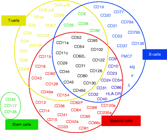
CD Antigens
Cell surface antigens of leukocytes are called CD antigens, and important for immune reactions of organisms. As lymphocytes mature, they express different protein receptors on the cell surface, which can aid in determining the type and maturation stage of the cells being examined. These proteins or antigen markers are called Clusters of Differentiation.
The term CD means a cluster of differentiation OR a cluster of determinants which indicates the lineage or maturational stage of lymphocytes. During the course of development from precursor cells into functionally mature forms, lymphocytes display a complex pattern of surface antigens, some of which are acquired at certain stages while others are lost.
These surface antigens were identified initially by monoclonal antibodies and the designations of the antibodies were used often as synonyms for the cell surface proteins they detected, giving rise to a plethora of different names. CD antigens are present on some subpopulations and functional types of leukocytes. CD antigens participate in immune reaction as receptors for cell communication (e.g. adherence molecules, antigen recognizing receptors).
CD antigen nomenclature describes different monoclonal antibodies from different sources that recognize identical antigens. Numbers are assigned arbitrarily. A small letter w before the number designation stands for "workshop". It indicates that the CD designation is tentative.
CD antigens are found on practically all known cell types. In some cases CD antigens are expressed only at certain stages of development or under certain conditions, for example after cell activation or in certain disease conditions. In Hematology the morphological criteria is for the description of specific developmental stages of lymphocytes unlike in CD antigens which the use of monoclonal antibodies allows the objective and precise analysis and standardized typing of mature and immature normal and malignant cells of all hematopoietic cell lineages. The use antibodies also helps to delineate the biologic traits that distinguish normal immune and hematopoietic cells from their malignant counterparts, which is utmost important in the understanding of hematological malignancies.
The expression of CD antigens is influenced by cytokines, such as binding of ligands to CD antigens which has shown to modulate the expression of cytokines. CD antigens have been shown to be identical with receptors of cytokines such as CD25 (TAC antigen).
CD antigens appear to carry out cytokine receptor-like functions such as CD27, CD30 and CD40. CD antigens are involved in modulating the biological activities of cytokines such as CD4, CD28 and CD40. CD antigens exist also in soluble forms for example CD14, CD21, CD23, CD27, CD100 and CD137.
The CD Antigen’s designation isn’t related to the biological function, thus CD antigens include receptors, glycans, adhesion molecules, membrane-bound enzymes, etc.
The most commonly know CD antigens are CD4 and CD8 which are markers for T-helper and T-suppressor cells, respectively. CD4 binds to relatively invariant sites on class II major histocompatibility complex molecules outside the peptide-binding groove, which interacts with the T-cell receptor. CD4 is also the central docking receptor for human immunodeficiency virus. CD8 binds to relatively invariant sites on class I major histocompatibility complex molecules outside the peptide-binding groove. CD8 is also expressed on a subset of dendritic cells. Other more important CD antigens include the leukocytes integrins (CD11/CD18) and the hematopoietic stem cell marker CD34.
CD69 is homologous to members of a supergene family of type II integral membrane proteins having C-type lectin domains. Although the precise functions of the CD-69 antigen is not known, evidence suggests that these proteins transmit mitogenic signals across the plasma membrane and are up- regulated in response to lymphocyte activation
In the last decade the wide palette of monoclonal antibodies has been prepared which recognise of CD antigens on human cells. Much less monoclonal antibodies are available specific for typing of domestic animal cells. CD antigens have been characterized as both transmembrane proteins and cell surface proteins anchored to the plasma membrane via covalent attachment to fatty acid-containing glycolipids such as glycosylphosphatidylinositol (GPI).


